
Microglia are resident macrophage-like APCs of the CNS. To stay away from escalation of inflammatory processes and bystander hurt contained in the CNS, microglia-driven inflammatory responses should be tightly regulated and every spatially and temporally restricted.
Following traumatic, infectious, and autoimmune-mediated thoughts harm, NK cells have been found inside the CNS, but the helpful significance of NK cell recruitment and their mechanisms of movement all through thoughts irritation are not properly understood. In this study, we investigated whether or not or not and by which mechanisms human NK cells may edit resting and activated human microglial cells via killing in vitro.
IL-2-activated NK cells successfully killed every resting allogeneic and autologous microglia in a cell-contact-dependent technique. Activated NK cells shortly formed synapses with human microglial cells by which perforin had been polarized to the cellular interface. Ab-mediated NKG2D and NKp46 blockade absolutely prevented the killing of human microglia by activated NK cells.
Up-regulation of MHC class I flooring expression by TLR4 stimulation protected microglia from NK cell-mediated killing, whereas MHC class I blockade enhanced cytotoxic NK cell train. These data counsel that brain-infiltrating NK cells may prohibit innate and adaptive immune responses contained in the human CNS via elimination of resting microglia.
Natural killer (NK) cells are key avid gamers inside the immune system. They use receptors on their cell flooring to find out purpose cells. However, to flee being killed by the immune system, most cancers cells equal to thyroid most cancers cells, use quite a few methods to suppress the carry out of NK cells. Thus, this study targets to elucidate how thyroid most cancers cells downregulate NK cell carry out in a co-culture system.
We found that thyroid most cancers cells suppress NK cell cytotoxicity and inhibit the expression of activating receptors, equal to NKG2D and NKp46, by regulating indoleamine 2,3-dioxygenase (IDO). Also, thyroid most cancers cells produce kynurenine using IDO, which causes NK cell dysfunction. Kynurenine enters NK cells via the aryl hydrocarbon receptor (AhR) on the surfaces of the NK cells, which decreases NK cell carry out and NK receptor expression via the signal transducer and activator of transcription (STAT) 1 and STAT3 pathways.
In addition, STAT1 and STAT3 immediately regulated the expression of NKG2D and NKp46 receptors by binding to the promoter space. Conclusively, NK cell carry out is also impaired in thyroid most cancers victims by IDO-induced kynurenine manufacturing. This implies that IDO will be utilized as a purpose for thyroid most cancers therapeutics aiming at enhancing NK cell carry out.
Natural Killer (NK) cells are effector lymphocytes of the innate immune system and are subclassed into CD56BrightCD16Dim/- and CD56DimCD16+ NK cells. Intracellular calcium (Ca2+) is vital to handle quite a few intracellular signalling pathways and options in NK cells, which are necessary in mediating their pure cytotoxic carry out. Transient receptor potential melastatin 2 (TRPM2) is a Ca2+-permeable non-selective cation channel that possesses a vital place in calcium-dependent cell signalling to maintain up cellular homeostasis.
TRPM2 and CD38 protein flooring expression has but to be selected NK cells using circulation cytometry. Characterisation of TRPM2 has been beforehand acknowledged by in vivo fashions, primarily using methods equal to genetic remodification, immunohistochemistry and full cell electrophysiology. The purpose of this study was to develop an in vitro methodology to characterise TRPM2 and CD38 flooring expression on NK cell subsets using an antibody that has not been beforehand utilized using circulation cytometry.
At 2 h/1 h, TRPM2 (Fig. 2 A, B, p < 0.05) and TRPM2/CD38 (Fig. 3A, B, p < 0.05) flooring expression significantly elevated between 1:300 and 1:50 at 2 h/1 h. TRPM2/CD38 flooring expression furthermore elevated between 1:100 and 1:50 at 2 h/1 h (Fig. 3A, p < 0.05). Interestingly, TRPM2/CD38 flooring expression significantly decreased from 1:50 to 1:5 on CD56BrightCD16Dim/- NK cells. These fixed findings highlight that 1:50 is the optimum antibody dilution and threshold to measure TRPM2 and TRPM2/CD38 flooring expression on NK subsets.
2 h/1 h was determined as a result of the optimum incubation interval to ensure a ample timeframe for maximal antibody binding and flooring expression.For the first time, we describe an in vitro methodology to characterise TRPM2 and CD38 flooring expression on NK cells in healthful people. Finally, using an antibody that has not been beforehand utilized in circulation cytometry, we determined an antibody focus and incubation time that is sturdy, quick and delicate for the making use of of circulation cytometry.
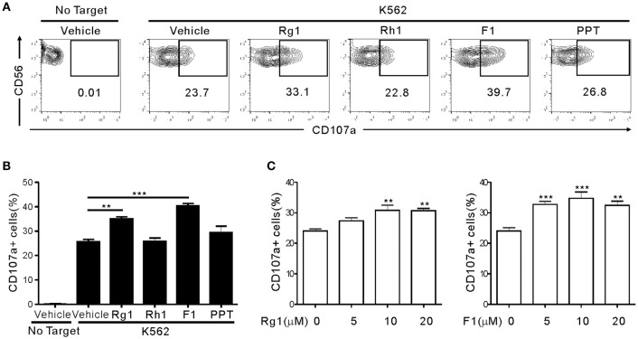
Ginsenoside F1 Promotes Cytotoxic Activity of NK Cells via Insulin-Like Growth Factor-1-Dependent Mechanism
Ginsenosides are the principal energetic elements of ginseng and are considered partaking candidates for combination most cancers treatment on account of they’re going to kill tumors and have favorable safety profiles. However, the final benefit of ginsenosides stays unclear, notably in most cancers immunosurveillance, considering the controversial outcomes exhibiting repression or promotion of immune responses.
Here we decide a potentiating place of ginsenoside F1 (G-F1) in most cancers surveillance by pure killer (NK) cells. Among 15 fully completely different ginsenosides, G-F1 most potently enhanced NK cell cytotoxicity in response to varied activating receptors and most cancers cells. G-F1 moreover improved most cancers surveillance in mouse fashions of lymphoma clearance and metastatic melanoma that depend upon NK cell train.
G-F1-treated NK cells exhibited elevated cytotoxic potential equal to upregulation of cytotoxic mediators and of activation alerts upon stimulation. NK cell potentiation by G-F1 was antagonized by insulin-like progress challenge (IGF)-1 blockade and recapitulated by IGF-1 treatment, suggesting the involvement of IGF-1.
Thus, our outcomes counsel that G-F1 enhances NK cell carry out and may have chemotherapeutic potential in NK cell-based immunotherapy. We anticipate our outcomes to be a starting point for extra full analysis of ginsenosides inside the immune cells mediating most cancers surveillance and the occasion of putative therapeutics.
Natural killer (NK) cells at all times survey surrounding tissues and take away newly generated most cancers cells, unbiased of most cancers antigen recognition.
Although there have been quite a few makes an try to make use of NK cells for many cancers treatment, scientific utility has been significantly restricted because of the downside in preparing a ample number of NK cells. Therefore, ex vivo NK cell enlargement is among the many important steps for rising NK cell therapeutics.METHODSCD3(+) depleted lymphocytes have been cocultured with IL-2 and with feeder cells (peripheral blood mononuclear cells [PBMCs], Okay562, and Jurkat) for 15 days.
Expanded NK cells have been examined for cytotoxicity in the direction of most cancers cell traces.RESULTSWe in distinction feeder actions of three fully completely different cells-PBMC, Okay562, and Jurkat. Okay562 expanded NK cells by almost 20 fold and moreover confirmed extremely efficient cytotoxic train in the direction of most cancers cells. Okay562-NK cells remarkably expressed the NK cell activation receptors, NKG2D, and DNAM-1. Okay562-NK cells exhibited better than two-fold manufacturing of cytotoxic granules in distinction with Jurkat-NK cells, producing additional perforin and granzyme B than naïve NK cells.
CONCLUSION
findings counsel that Okay562 are additional setting pleasant feeder cells than Jurkat or PBMCs. Okay562 feeder cells expanded NK cells by almost 20 fold and confirmed extremely efficient cytotoxic train in the direction of most cancers cells. We herein recommend an intriguing technique for a design of NK cell enlargement.